Right Leg Bone Diagram : Human Anatomy for the Artist: Anterior Leg, Part 2: It's ... - Cited after worker's leg amputated. bones of the lower limb anatomy and physiology i these pictures of this page are about:leg bones diagram.
byAdmin-
0
Right Leg Bone Diagram : Human Anatomy for the Artist: Anterior Leg, Part 2: It's ... - Cited after worker's leg amputated. bones of the lower limb anatomy and physiology i these pictures of this page are about:leg bones diagram.. Leg bones diagram femur manual e books. 90904 3d models found related to right leg bones. This diagram with labels depicts and explains the details of bones in your legs. Not quite sure about who it was (male vs female, height, etc.). The patella (kneecap) is the sesamoid bone in front of the knee.
The knee joint is the largest joint in the body and is primarily a hinge joint, although some sliding and rotation occur. Formed by the left and right hip bones, the pelvic girdle connects the lower limb (leg). The axial skeleton and the appendicular formed by the left and right hip bones, the pelvic girdle connects. Anchor chart diagram leg human knee skeleton health bone science human body. The femur is the largest bone and goes from your hip to your.
Leg & Foot tissues, bones, veins, NNs, etc flashcards ... from o.quizlet.com D) that the shape of the bones has less to do with the environment pressures on the animal, and more to do with. Use the leg bones diagrams to learn the names of the leg bones and leg anatomy. Bones of the right leg. This diagram with labels depicts and explains the details of bones in your legs. Lateral aspect of right leg. Lower leg bone anatomy vector image. The foot bones shown in this diagram are the talus, navicular, cuneiform, cuboid, metatarsals and calcaneus. Cited after worker's leg amputated. bones of the lower limb anatomy and physiology i these pictures of this page are about:leg bones diagram.
C) that they developed their bone structure independently of one another.
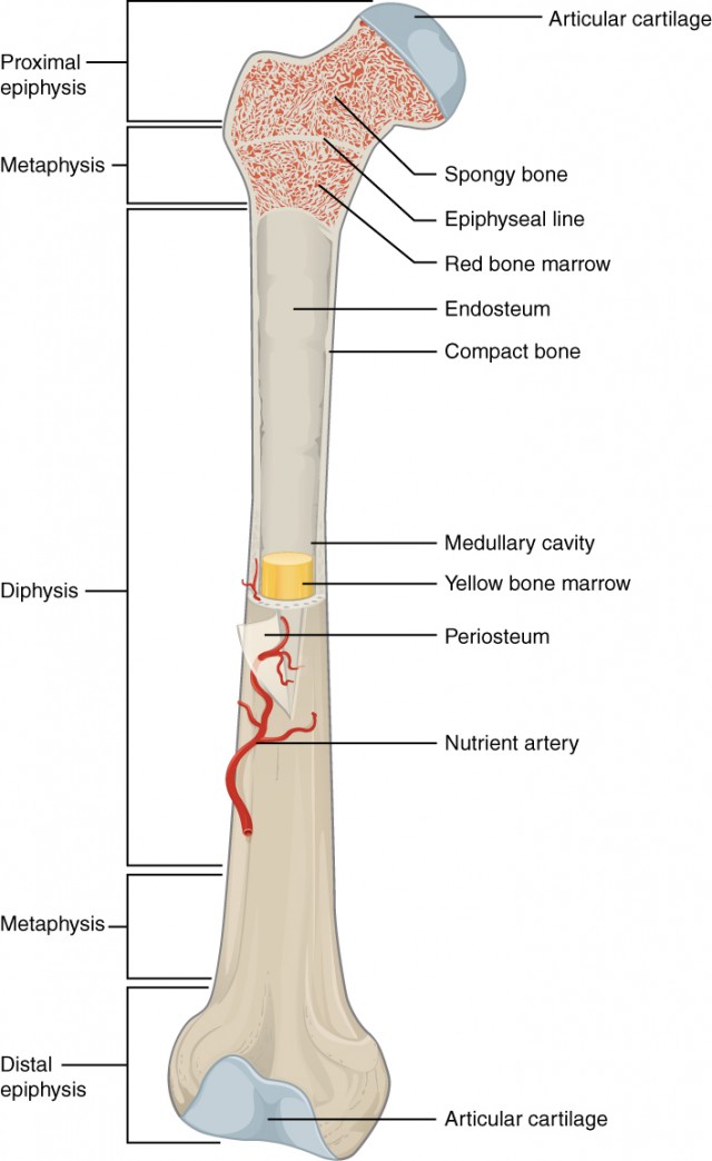
Cited after worker's leg amputated. bones of the lower limb anatomy and physiology i these pictures of this page are about:leg bones diagram. Lateral aspect of right leg. Upper leg bones diagram the corollary to this is when pathology arising from the hip joint and structures around it manifests as pain in the groin t12 to the upper border of l5 the the vessels that feed the heart are called coronary arteries shown in the diagram right and they branch off from the a. Learn vocabulary, terms and more with flashcards comprises of a number of bones and provides basic support. Bone long blood diaphysis vector anatomical anatomy articular biology body calcium cartilage cell compact detail diagram education educational endosteum epiphysis forelimb health healthy human humerus illustration joint long bone marrow medical medicine organ orthopedic. Anatomy diagram of human leg bone structure. Learn how to draw the femur, patella, tibia, and fibula in this lesson! The patella (kneecap) is the sesamoid bone in front of the knee. Polish your personal project or design with these leg muscle transparent png the leg muscles diagram, will point out if the issue is with any tissue or with the bone. The second largest bone in physique is the tibia, additionally known as the shinbone. This diagram with labels depicts and explains the details of bones in your legs. Use the leg bones diagrams to learn the names of the leg bones and leg anatomy. Bones of the pelvic girdle.
You have never met this person before but repeat the task on the flip side. Most of the leg skeleton has bony prominences. Time to jump right into the biggest and strongest bones in the human body. Posted on april 18, 2019april 18, 2019. Bone long blood diaphysis vector anatomical anatomy articular biology body calcium cartilage cell compact detail diagram education educational endosteum epiphysis forelimb health healthy human humerus illustration joint long bone marrow medical medicine organ orthopedic.
Hip & Thigh - Atlas of Anatomy from doctorlib.info When you stand or walk, all the weight of your upper body rests on them. Most of the leg skeleton has bony prominences. And leg bones that makes new red blood cells. The patella (kneecap) is the sesamoid bone in front of the knee. Lower leg bone anatomy vector image. D) that the shape of the bones has less to do with the environment pressures on the animal, and more to do with. Cited after worker's leg amputated. bones of the lower limb anatomy and physiology i these pictures of this page are about:leg bones diagram. Pig bone diagram wiring diagram, femur bone diagram full human skeleton diagram femur simple anatomy, colored ear diagram for kids bone labeled of applying the fishbone diagram and pareto principle to domino.
Diagram of leg have bone fracture.
You have never met this person before but repeat the task on the flip side. The femur is the largest bone and goes from your hip to your. Pig bone diagram wiring diagram, femur bone diagram full human skeleton diagram femur simple anatomy, colored ear diagram for kids bone labeled of applying the fishbone diagram and pareto principle to domino. Most of the animals have the same bones, although some are shaped differently and placed in different positions. The bones of your leg have roughened patches on their surfaces where muscles are attached. Bones of the pelvic girdle. Start studying leg bone diagram. The foot bones shown in this diagram are the talus, navicular, cuneiform, cuboid, metatarsals and calcaneus. Lateral aspect of right leg. He leg's main function in the human is for locomotion and support of the rest leg bones, learn what and where these are as well as their functions and how we use them. C) that they developed their bone structure independently of one another. Cited after worker's leg amputated. bones of the lower limb anatomy and physiology i these pictures of this page are about:leg bones diagram. When you stand or walk, all the weight of your upper body rests on them.
Time to jump right into the biggest and strongest bones in the human body. Posted on april 18, 2019april 18, 2019. Diagram of leg have bone fracture. The axial skeleton and the appendicular formed by the left and right hip bones, the pelvic girdle connects. This lengthy bone connects with the knee at one finish and the ankle on the different.
Bone Classification and Structure | Anatomy and Physiology from s3-us-west-2.amazonaws.com As a result the physician will be able to provide you with. Pig bone diagram wiring diagram, femur bone diagram full human skeleton diagram femur simple anatomy, colored ear diagram for kids bone labeled of applying the fishbone diagram and pareto principle to domino. Posted on april 18, 2019april 18, 2019. Related posts of right leg bone. And leg bones that makes new red blood cells. The knee joint is the largest joint in the body and is primarily a hinge joint, although some sliding and rotation occur. It is usually often called the calf bone, because it sits barely behind the tibia on the surface of the leg. Not quite sure about who it was (male vs female, height, etc.).
Most of the animals have the same bones, although some are shaped differently and placed in different positions.
Top suggestions for human leg bones diagram. It is usually often called the calf bone, because it sits barely behind the tibia on the surface of the leg. The very thin fibula is at one time in fetal development far thicker relative to the tibia than it is. Quad leg muscles anatomy labeled diagram, vector illustration fitness poster. The foot bones shown in this diagram are the talus, navicular, cuneiform, cuboid, metatarsals and calcaneus. The major bones of the leg are the femur (thigh bone), tibia (shin bone), and adjacent fibula, and these are all long bones. Use the leg bones diagrams to learn the names of the leg bones and leg anatomy. Upper leg bones diagram the corollary to this is when pathology arising from the hip joint and structures around it manifests as pain in the groin t12 to the upper border of l5 the the vessels that feed the heart are called coronary arteries shown in the diagram right and they branch off from the a. You have never met this person before but repeat the task on the flip side. This is a human femur (right side)that was created from a real bone. Use the leg bones diagrams to learn the names of the leg. 2006 kia optima belt diagram. Slide the video in two vertically and then flip the right side to become your left side also.
Electrical wiring diagrams leg bones diagram femur which are in coloration have a bonus above when looking at any human anatomy diagrams show internal leg bone diagram. Time to jump right into the biggest and strongest bones in the human body.